Radiology & Imaging Services
Other Programs & Services
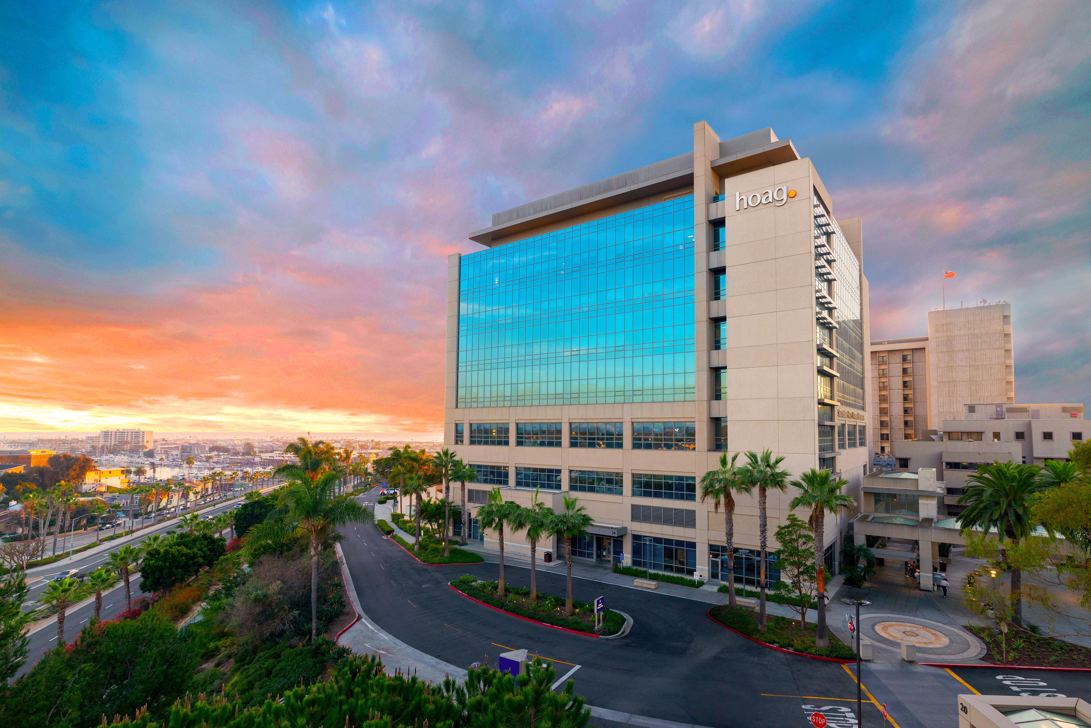
1 Hoag Dr, Newport Beach, CA 92663
(949) 764-4624
- About
- Services
- Appointments
- Meet the Team
- More Info
- Locations
Unlike many other Radiology/Imaging centers, Hoag hospitals and outpatient centers meet the strict requirements set by the DNV NIAHO Accreditations Program.
Our Radiologists
A Higher Standard
Our Leadership Team
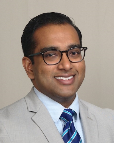
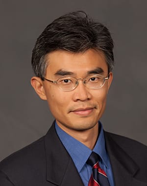
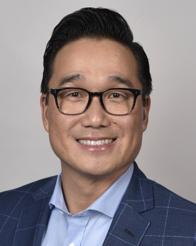
Yingding Xu, MD
<p>Dr. Xu joined NHRA in 2019. He graduated with honor from California Institute of Technology and completed his medical school training at Stanford University School of Medicine. He was the recipient of the Resident Teaching Award during his internship year at Kaiser Permanente Santa Clara. He then returned to Stanford Hospital and Clinics for his radiology residency and fellowship where he received the Roentgen Resident Research Award from RSNA and served as the chief fellow of the musculoskeletal imaging fellowship. Dr. Xu also completed fellowship-level training in breast imaging at Stanford University.</p>
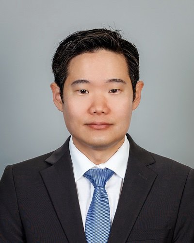
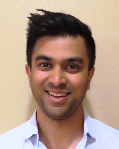
Tust Techasith, MD
<p>Dr. Tust Techasith specializes in Vascular and Interventional Radiology, with particular emphasis on minimally invasive treatment of cancer and venous disease.</p> <p>Dr. Techasith graduated from Harvard Medical School in 2011. He completed his internship at Santa Clara Valley Medical Center where he was awarded Intern of the Year. He subsequently completed both his residency training in Diagnostic Radiology, serving as chief resident, and fellowship training in Vascular and Interventional Radiology at Stanford University Medical Center.</p> <p>Dr. Techasith has authored or co-authored over 30 scientific abstracts, book chapters, and peer-reviewed publications in the field of diagnostic and interventional radiology. He has also been an invited lecturer at various residency training programs, universities, and national meetings.</p>
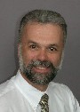
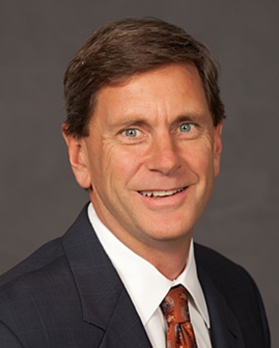
Humberto Wong, MD
<p>Dr. Wong joined the radiology department at Hoag in 2008. He completed all of his medical training at Stanford University Medical Center including his medical degree, radiology residency and cardiovascular imaging fellowship. He has extensive experience in cardiac MRI and cardiac/coronary CTA as well as interpretation of CT angiography for valvular procedure such as TAVR. He is co-President of Newport Harbor Radiology Associates and Adjunct Clinical Instructor in the Cardiovascular Imaging in the Department of Radiology at Stanford University Medical Center.</p>
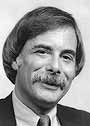
Roger H. Thomas, MD
<p>Dr. Roger Thomas joined NHRA in 1975. Dr. Thomas received honors with Alpha Omega Alpha National Society. He is a Fellow in the American College of Radiology.</p>

Christopher B. Baker, MD
<p>Dr. Baker specializes in the diagnosis and treatment of vascular diseases of the brain and spine. He has been in practice since finishing his training at Stanford University in 2009, and joined Newport Harbor Radiology in 2017. After graduating from Brigham Young University with a Bachelor of Science in Molecular Biology he attended New Jersey Medical School. He then went on to residency in Radiology at Maine Medical Center and completed fellowships in Vascular and Interventional Radiology and Interventional Neuroradiology. He has an academic appointment as Adjunct Clinical Associate Professor at Stanford University School of Medicine and is routinely invited to speak on endovascular treatment of acute stroke at the Annual Scientific Meeting of the Society of Interventional Radiology. He has also participated in several international clinical trials studying the endovascular treatment of brain aneurysms, acute stroke, and carotid artery disease.</p>

Jennifer M. Overstreet, MD
Associate Director of Breast Imaging
Radiology & Imaging Services
Your care starts here
1
Find the right provider
Get care from medical providers that fit your needs in a location near you.
Find a provider2
3
Get in touch
Didn’t see what you’re looking for? Reach out and we’ll make sure you get what you need.
Contact usServices
In MR arthrography, a small amount of contrast (gadolinium) is injected directly into the joint of interest (e.g. hip, shoulder, knee). This allows for detailed evaluation of structures that are difficult to see by conventional MRI examination. Structures particularly well evaluated by this technique include the joint capsule, ligaments, tendons and labrum.
In addition to the standard MRI examination routinely used to image the brain and central nervous system, a number of advanced imaging techniques are available which are helpful in the diagnosis and management of various neurological conditions, including brain tumors, stroke, and dementia. These techniques include Magnetic Resonance (MR) Angiography, MR Spectroscopy, MR Perfusion, Functional MRI, and Diffusion Tensor Imaging (DTI) and Tractography.
A biopsy is a procedure in which cells are removed from suspicious areas and analyzed by a physician specializing in pathology, to determine whether or not cancer is present. Because suspicious areas in the breast are more commonly discovered by imaging rather than by being felt in an external examination, imaging is often the best method to direct the breast biopsy procedure. Biopsies done under radiologic guidance are performed with a needle instead of subjecting the patient to disfiguring surgery.
Magnetic resonance imaging (MRI) of the breast is an important new approach in which to diagnose and characterize abnormalities in the breasts. Because images produced by MRI are very detailed, this technology can detect small changes or abnormalities.
Breast MRI frequently can be used to determine whether or not an area is cancerous, thereby avoiding unnecessary biopsies; it is especially helpful for evaluation of very dense breasts. This exam is also used to check for leakage or rupture of breast implants.
Breast ultrasound examination produces images of your breast using inaudible sound waves in a frequency range far above the range of human hearing. This procedure also is known as a sonogram. A breast ultrasound may be ordered by your physician or recommended by your radiologist to obtain more information about a possible abnormality. It is frequently done to assess the characteristics of a lump.
Magnetic Resonance Imaging (MRI) produces images of the body’s internal structures by passing radio waves through a powerful magnetic field. Differing frequencies of radio waves are produced by the different body structures, in return, and these are mapped and converted into digital images by a computer. MRI is especially good for imaging soft tissues in the body, including the brain, nerves, muscles and organs.
Detailed MR images allow physicians to better evaluate various parts of the body and certain diseases that may not be assessed adequately with other imaging methods such as x-ray, ultrasound or computed tomography (also called CT or CAT scanning).
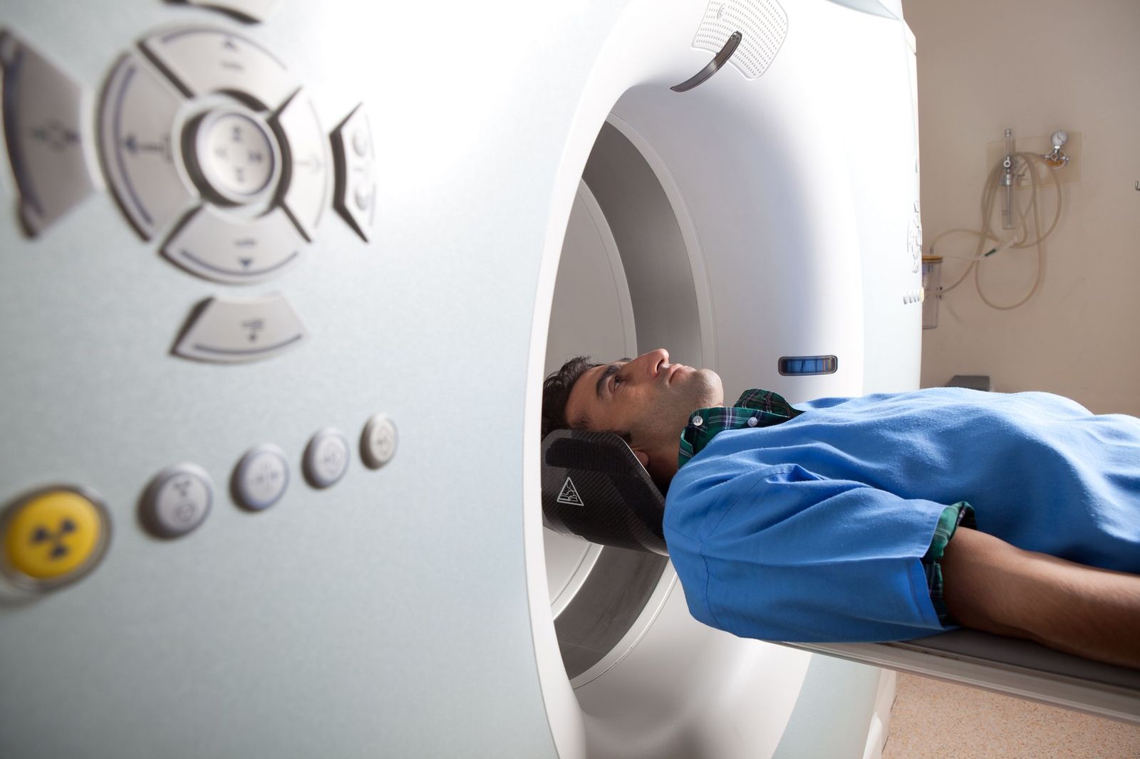
CT scanning – sometimes called CAT scanning – is a noninvasive medical test that helps physicians diagnose and treat medical conditions.
CT scanning combines special X-ray equipment with sophisticated computers to produce multiple images or pictures of the inside of the body.
Coronary CT angiography (CTA) is a non-invasive examination used to visualize the coronary arteries in order to assess whether there are blockages or restrictions to the flow of blood (and oxygen) to the heart. Coronary CTA is a form of CT (computed tomography), which uses the latest state of the art CT scanners in order to freeze cardiac motion while contrast is administered through an IV in the arm, and allow for both 2D, 3D and 4D visualization of cardiac structures such as the coronary arteries.
Dual-energy X-ray absorptiometry (DEXA) is a technology that uses a very low amount of X-ray energy to detect the presence of osteoporosis. Bone mineral content tends to begin declining in people who are in their mid-30s or older, and the loss of bone tissue accelerates in women after menopause. Reduction of mineral content, characterized by porous and brittle bones, is known as osteoporosis. It’s a potentially serious condition because it makes bones more vulnerable to fractures and ultimately can lead to diminished mobility and independence.
DEXA scanning can identify low bone density in patients at an early stage, enabling doctors to prescribe appropriate treatment before the condition worsens. Images of the lower spine and hips are most often used in checking for osteoporosis.
Some internal organs—such as kidneys, blood vessels and the organs of the gastrointestinal tract—can best be visualized on X-ray film when the patient ingests a contrast material. This blocks nearly all photons and makes tissues in which the contrast is distributed appear bright white. The contrast medium may be given orally, intravenously or rectally, depending upon the area to be studied.
Interventional radiology constitutes a major advance in medicine, encompassing procedures that eliminate the need for large incisions, with far less risk, less pain and shorter recovery times than traditional surgical procedures. NHRA has a staff of fellowship-trained interventional radiologists who specialize in neurological (head and spine) and body procedures.
Interventional neuroradiology (also known as neurointerventional surgery, or endovascular neurosurgery, or endovascular surgical neuroradiology) is a subspecialty of Hoag Radiology that utilizes advanced imaging and minimally invasive techniques for the treatment of life-threatening conditions of the blood vessels and of the brain and spine, such as acute stroke, cerebral aneurysms, arterial stenosis, and vascular malformations.
A mammogram is a low-dose X-ray examination of the breast tissue. Mammograms are commonly performed to look for breast cancer, but also can detect non-cancerous masses, cysts, calcifications and sometimes breast implant ruptures. Traditional analog mammograms are recorded and stored on X-ray film. With digital mammography, the images are recorded and stored on computerized media. The method of performing the exam and the image quality are identical for analog and digital mammography.
Hoag is proud to announce that it is the first hospital on the West Coast to routinely offer patients a revolutionary hybrid technology that will forever change the way neurology, cancer and cardiac patients are diagnosed and treated.
Positron emission tomography/magnetic resonance imaging (MR/PET), is an innovative imaging technology that will help in early and accurate diagnosis of various cancers, degenerative neurological disorders such as Alzheimer’s disease, mild traumatic brain injury and heart disease. MR/PET will also help physicians tailor treatment options to ensure patients receive the care that will most likely work best for them.
Magnetic Resonance Imaging is truly one of the medical wonders of the modern world, giving physicians the ability to see internal tissues and even brain function in three-dimensional detail.
Hoag stands ready, with a convenient MRI imaging center near you. From joint issues to next-generation techniques like functional magnetic resonance imaging (fMRI) and Magnetic Resonance Venography (MRV), Hoag has the tools and technology to get to the root of the problem and find the treatment you need to heal.
Nuclear Medicine is a subspecialty within the field of radiology. It includes diagnostic imaging studies that demonstrate body anatomy and function. The images are based on the distribution of a radioactive substance given to the patient, either intravenously, by mouth or inhaled into the lungs. Generally, radiation to the patient is similar to that resulting from standard x-ray examinations. Nuclear medicine images can assist the physician in diagnosing diseases. Tumors, infections and other disorders can be diagnosed by evaluation organ function.
Nuclear Medicine is a subspecialty within the field of radiology. It includes diagnostic imaging studies that demonstrate body anatomy and function. The images are based on the distribution of a radioactive substance given to the patient, either intravenously, by mouth or inhaled into the lungs. Generally, radiation to the patient is similar to that resulting from standard x-ray examinations. Nuclear medicine images can assist the physician in diagnosing diseases. Tumors, infections and other disorders can be diagnosed by evaluation organ function.
Positron Emission Tomography, also called PET, is a diagnostic tool that produces images that demonstrate organ function. PET images are based on the detection of subatomic particles, specifically positrons.
Positrons are emitted from a radioactive substance administered to the patient and are ultimately detected by special crystals within the PET scanner. This information is then digitized to produce a 3-Dimensional image of the whole body or of a specific organ. With PET/CT, Computerized Tomography images are also obtained, which show detailed views of the structure of the body part being examined. The two types of images are “fused” by a computer into a set of pictures that shows both anatomical detail and function of the area being examined.
Spinal injections are performed either to confirm a suspected diagnosis or to reduce pain and inflammation resulting from spinal problems. These injections are performed under fluoroscopy by an interventional radiologist.
Game-changing medical innovations are being pushed to the forefront every day, and in few areas is that more true than medical imaging, including advancements in ultrasound technology.
Hoag is at the forefront of these groundbreaking techniques. From monitoring fetal development and allowing parents to see their child’s face long before birth, to doppler ultrasound techniques that give cardiologists a way to visualize blood flow through the heart and blood vessels as it happens, Hoag is leading the way.
Select an Imaging and Radiology Service
Online Booking
Book by Phone
In MR arthrography, a small amount of contrast (gadolinium) is injected directly into the joint of interest (e.g. hip, shoulder, knee). This allows for detailed evaluation of structures that are difficult to see by conventional MRI examination. Structures particularly well evaluated by this technique include the joint capsule, ligaments, tendons and labrum.
In addition to the standard MRI examination routinely used to image the brain and central nervous system, a number of advanced imaging techniques are available which are helpful in the diagnosis and management of various neurological conditions, including brain tumors, stroke, and dementia. These techniques include Magnetic Resonance (MR) Angiography, MR Spectroscopy, MR Perfusion, Functional MRI, and Diffusion Tensor Imaging (DTI) and Tractography.
A biopsy is a procedure in which cells are removed from suspicious areas and analyzed by a physician specializing in pathology, to determine whether or not cancer is present. Because suspicious areas in the breast are more commonly discovered by imaging rather than by being felt in an external examination, imaging is often the best method to direct the breast biopsy procedure. Biopsies done under radiologic guidance are performed with a needle instead of subjecting the patient to disfiguring surgery.
Magnetic resonance imaging (MRI) of the breast is an important new approach in which to diagnose and characterize abnormalities in the breasts. Because images produced by MRI are very detailed, this technology can detect small changes or abnormalities.
Breast MRI frequently can be used to determine whether or not an area is cancerous, thereby avoiding unnecessary biopsies; it is especially helpful for evaluation of very dense breasts. This exam is also used to check for leakage or rupture of breast implants.
Breast ultrasound examination produces images of your breast using inaudible sound waves in a frequency range far above the range of human hearing. This procedure also is known as a sonogram. A breast ultrasound may be ordered by your physician or recommended by your radiologist to obtain more information about a possible abnormality. It is frequently done to assess the characteristics of a lump.
Magnetic Resonance Imaging (MRI) produces images of the body’s internal structures by passing radio waves through a powerful magnetic field. Differing frequencies of radio waves are produced by the different body structures, in return, and these are mapped and converted into digital images by a computer. MRI is especially good for imaging soft tissues in the body, including the brain, nerves, muscles and organs.
Detailed MR images allow physicians to better evaluate various parts of the body and certain diseases that may not be assessed adequately with other imaging methods such as x-ray, ultrasound or computed tomography (also called CT or CAT scanning).
CT scanning – sometimes called CAT scanning – is a noninvasive medical test that helps physicians diagnose and treat medical conditions.
CT scanning combines special X-ray equipment with sophisticated computers to produce multiple images or pictures of the inside of the body.
Coronary CT angiography (CTA) is a non-invasive examination used to visualize the coronary arteries in order to assess whether there are blockages or restrictions to the flow of blood (and oxygen) to the heart. Coronary CTA is a form of CT (computed tomography), which uses the latest state of the art CT scanners in order to freeze cardiac motion while contrast is administered through an IV in the arm, and allow for both 2D, 3D and 4D visualization of cardiac structures such as the coronary arteries.
Some internal organs—such as kidneys, blood vessels and the organs of the gastrointestinal tract—can best be visualized on X-ray film when the patient ingests a contrast material. This blocks nearly all photons and makes tissues in which the contrast is distributed appear bright white. The contrast medium may be given orally, intravenously or rectally, depending upon the area to be studied.
Interventional radiology constitutes a major advance in medicine, encompassing procedures that eliminate the need for large incisions, with far less risk, less pain and shorter recovery times than traditional surgical procedures. NHRA has a staff of fellowship-trained interventional radiologists who specialize in neurological (head and spine) and body procedures.
Hoag is proud to announce that it is the first hospital on the West Coast to routinely offer patients a revolutionary hybrid technology that will forever change the way neurology, cancer and cardiac patients are diagnosed and treated.
Positron emission tomography/magnetic resonance imaging (MR/PET), is an innovative imaging technology that will help in early and accurate diagnosis of various cancers, degenerative neurological disorders such as Alzheimer’s disease, mild traumatic brain injury and heart disease. MR/PET will also help physicians tailor treatment options to ensure patients receive the care that will most likely work best for them.
Magnetic Resonance Imaging is truly one of the medical wonders of the modern world, giving physicians the ability to see internal tissues and even brain function in three-dimensional detail.
Hoag stands ready, with a convenient MRI imaging center near you. From joint issues to next-generation techniques like functional magnetic resonance imaging (fMRI) and Magnetic Resonance Venography (MRV), Hoag has the tools and technology to get to the root of the problem and find the treatment you need to heal.
Nuclear Medicine is a subspecialty within the field of radiology. It includes diagnostic imaging studies that demonstrate body anatomy and function. The images are based on the distribution of a radioactive substance given to the patient, either intravenously, by mouth or inhaled into the lungs. Generally, radiation to the patient is similar to that resulting from standard x-ray examinations. Nuclear medicine images can assist the physician in diagnosing diseases. Tumors, infections and other disorders can be diagnosed by evaluation organ function.
Nuclear Medicine is a subspecialty within the field of radiology. It includes diagnostic imaging studies that demonstrate body anatomy and function. The images are based on the distribution of a radioactive substance given to the patient, either intravenously, by mouth or inhaled into the lungs. Generally, radiation to the patient is similar to that resulting from standard x-ray examinations. Nuclear medicine images can assist the physician in diagnosing diseases. Tumors, infections and other disorders can be diagnosed by evaluation organ function.
Positron Emission Tomography, also called PET, is a diagnostic tool that produces images that demonstrate organ function. PET images are based on the detection of subatomic particles, specifically positrons.
Positrons are emitted from a radioactive substance administered to the patient and are ultimately detected by special crystals within the PET scanner. This information is then digitized to produce a 3-Dimensional image of the whole body or of a specific organ. With PET/CT, Computerized Tomography images are also obtained, which show detailed views of the structure of the body part being examined. The two types of images are “fused” by a computer into a set of pictures that shows both anatomical detail and function of the area being examined.
Spinal injections are performed either to confirm a suspected diagnosis or to reduce pain and inflammation resulting from spinal problems. These injections are performed under fluoroscopy by an interventional radiologist.
Meet The Team
Meet the Team
Gerard K. Nguyen, MD
Radiology

Trushar Patel, MD
<p>Dr. Trushar Patel joined NHRA in 2014. He specializes in Vascular and Interventional Radiology with a focus in Interventional Oncology and Venous Disease. After finishing residency at Cornell Medical Center with additional training at Memorial Sloan Kettering Cancer Center, Dr. Patel completed a fellowship in Vascular and Interventional Radiology at the University of California, San Francisco.</p>
John G. Lieu, MD
<p>Dr. John Lieu Joined NHRA in 2009. Prior to NHRA, he was a Clinical Instructor in the Department of Radiology at Stanford.</p>
Avinash Mesipam, MD
<p>Dr. Avinash Mesipam joined NHRA in 2020. He specializes in the diagnosis and treatment of vascular diseases of the brain and spine.</p> <p>Dr. Mesipam graduated from Loma Linda School of Medicine in 2012. He completed his internship at Cottage Hospital in Santa Barbara. He subsequently completed his residency in Diagnostic Radiology at Loma Linda University.</p> <p>After completion of residency, Dr. Mesipam completed a fellowship in Vascular and Interventional Radiology at Brown University in 2018. He then completed a fellowship in Diagnostic Neuroradiology at Loma Linda University in 2019, this was followed by a fellowship in Endovascular Surgical Neuroradiology at the Mallinckrodt Institute of Radiology at Washington University School of Medicine in St. Louis in 2020.</p>
Shaya Ghazinoor, MD
<p>Dr. Shaya Ghazinoor joined NHRA in 2013. She graduated summa cum laude from UCLA and went on to receive her medical degree from University of California, San Francisco. She completed her radiology residency at Stanford and went on to subspecialize in Sports Imaging with a 1 year musculoskeletal MRI fellowship. Prior to joining NHRA, Dr. Ghazinoor practiced in an outpatient setting in Orange County.</p>
Winston S. Whitney, MD
<p>Dr. Winston Whitney joined NHRA in 1993. Dr. Whitney is currently the Medical Director of Hoag-Huntington Beach Imaging Center and the Medical Director of Hoag Hospital Outpatient Imaging. He has served as the Director of Radiology Quality Assurance, Director of NHRA Radiologist Recruitment, and as a member of the Hoag Radiology Executive Committee, Hoag Cancer Committee and Hoag Lung Cancer Committee.</p>
Wilson Lin, MD
<p>Dr. Wilson Lin joined NHRA in 2019. He graduated from the University of California Los Angeles (Bachelors of Science – Neuroscience) and University of California San Francisco (Medical Degree, with distinction). While in medical school at the University of California San Francisco, Dr. Lin completed an additional yearlong Clinical and Translational Research Fellowship funded by the NIH, during which he investigated imaging biomarkers for cartilage degeneration. During his residency at Stanford Hospital and Clinics, Dr. Lin was awarded the Radiological Society of North America Research Trainee Prize, which selected one resident nationwide for excellence in research. Dr. Lin specializes in musculoskeletal radiology. Dr. Lin has also spent several years working on the Google Brain Project, specifically on the role of artificial intelligence in radiology.</p>
Michael N. Brant-Zawadzki, MD
Michael Brant-Zawadzki, M.D., F.A.C.R. is currently the Vice President of Clinical Research Administration, Center for Accelerating Technology and Life Sciences Translation (CATALiST), and Addiction Services at Hoag Hospital. Formerly, he was Hoag’s Senior Physician Executive, facilitating business and strategic relationships with specialist clinicians, overseeing a platform for programmatic care that integrates specialized services with population health. He also served as Executive Medical Director of the Pickup Family Neurosciences Institute, where he held the Sandi and Ron Simon Endowed Chair, helping Hoag transform health care strategy, focusing it on specific health conditions using a program-driven, physician-led, multidisciplinary team approach, measured by patient-focused outcomes. Dr. Brant-Zawadzki has a BA from Stanford, and earned his MD from the University of Cincinnati, graduating first in his class. He trained in diagnostic radiology and in interventional neuroradiology at Stanford. He spent six years in the Department of Radiology at UCSF; he also held an Adjunct Professorship at Stanford until 2023. Dr. Brant-Zawadzki has authored over 250 papers and numerous textbooks, serves on a variety of editorial boards for professional journals, and has served on numerous professional organization boards and committees. He lectures internationally. He is a Fellow of the American College of Radiology and a Gold Medal recipient from the Society of Magnetic Resonance in Medicine and the American Society of Neuroradiology. He earned the Certificate in Leadership for Health Care Transformation from UC Irvine's Paul Merage School of Business. He also is a founder of, and consultant to, a number of companies in the biotech and radiology fields.
Michael C. Jung, MD
<p>Michael Jung joined NHRA in 2011. Dr. Jung graduated Phi Beta Kappa with Distinction from Stanford University and completed his medical school training at the University of Pennsylvania. Following residency training in diagnostic radiology at the University of California-San Diego, he completed a two-year fellowship in diagnostic neuroradiology at the Massachusetts General Hospital, Harvard Medical School. Dr. Jung is certified by the American Board of Radiology and holds a Certificate of Added Qualification in Neuroradiology.</p>

Scott T. Williams, MD
<p>Dr. Williams has been a member of Newport Harbor Radiology Associates since 2006. He joined NHRA after completing his diagnostic radiology residency and subsequent body imaging fellowship at Stanford. His Stanford training included advanced body imaging techniques, including cardiac and cardiovascular imaging, as well as image guided interventional procedures.</p> <p>Dr. Williams currently serves as the Chief of Service for the Department of Radiology, Hoag Hospital Newport Beach, and surrounding centers. He is the Radiology Supervisor for Hoag Urgent Care centers located throughout Orange County. Since 2008 he has served as Section Chief for Cardiovascular Imaging. He currently serves as the Director of the 3D Imaging Lab and is the founder of the 3D printing program at Hoag. He is an integral member of the Trans Arterial Valve Replacement (TAVR) team for the Jeffrey M. Carlton Heart and Vascular Institute. Dr. Williams is a founding member and physician champion of the Hoag Physician Wellness Committee. He also serves as a member of the Quality Improvement Committee of the Board for Hoag Hospital.</p>
Yingding Xu, MD
<p>Dr. Xu joined NHRA in 2019. He graduated with honor from California Institute of Technology and completed his medical school training at Stanford University School of Medicine. He was the recipient of the Resident Teaching Award during his internship year at Kaiser Permanente Santa Clara. He then returned to Stanford Hospital and Clinics for his radiology residency and fellowship where he received the Roentgen Resident Research Award from RSNA and served as the chief fellow of the musculoskeletal imaging fellowship. Dr. Xu also completed fellowship-level training in breast imaging at Stanford University.</p>
Karence K. Chan, MD
<p>Dr. Karence Chan joined NHRA in 1998. Dr. Chan was a Clinical Instructor in the Radiology Department at the University of California, San Diego Medical Center from 1998-2000.</p>
Tust Techasith, MD
<p>Dr. Tust Techasith specializes in Vascular and Interventional Radiology, with particular emphasis on minimally invasive treatment of cancer and venous disease.</p> <p>Dr. Techasith graduated from Harvard Medical School in 2011. He completed his internship at Santa Clara Valley Medical Center where he was awarded Intern of the Year. He subsequently completed both his residency training in Diagnostic Radiology, serving as chief resident, and fellowship training in Vascular and Interventional Radiology at Stanford University Medical Center.</p> <p>Dr. Techasith has authored or co-authored over 30 scientific abstracts, book chapters, and peer-reviewed publications in the field of diagnostic and interventional radiology. He has also been an invited lecturer at various residency training programs, universities, and national meetings.</p>
Thomas E. Velling, MD
<p>Dr. Thomas Velling joined NHRA in 2001. He currently serves as Director of the Interventional Radiology Service at Hoag Hospital in Newport Beach.</p>
Mark Z. Chen, MD
Radiology
Frederick A. Birnberg, MD
<p>Dr. Frederick Birnberg joined NHRA in 1988. He was a former Assistant Professor in the Department of Radiology at the University of Michigan where he received Outstanding Faculty Teacher of the Year Award. Now, Dr. Birnberg is a clinical professor of Radiology at the University of California Davis. Prior to joining Newport Harbor Radiology he was Chief of Radiology for Kaiser, Sacramento.</p>
Humberto Wong, MD
<p>Dr. Wong joined the radiology department at Hoag in 2008. He completed all of his medical training at Stanford University Medical Center including his medical degree, radiology residency and cardiovascular imaging fellowship. He has extensive experience in cardiac MRI and cardiac/coronary CTA as well as interpretation of CT angiography for valvular procedure such as TAVR. He is co-President of Newport Harbor Radiology Associates and Adjunct Clinical Instructor in the Cardiovascular Imaging in the Department of Radiology at Stanford University Medical Center.</p>
Christopher B. Baker, MD
<p>Dr. Baker specializes in the diagnosis and treatment of vascular diseases of the brain and spine. He has been in practice since finishing his training at Stanford University in 2009, and joined Newport Harbor Radiology in 2017. After graduating from Brigham Young University with a Bachelor of Science in Molecular Biology he attended New Jersey Medical School. He then went on to residency in Radiology at Maine Medical Center and completed fellowships in Vascular and Interventional Radiology and Interventional Neuroradiology. He has an academic appointment as Adjunct Clinical Associate Professor at Stanford University School of Medicine and is routinely invited to speak on endovascular treatment of acute stroke at the Annual Scientific Meeting of the Society of Interventional Radiology. He has also participated in several international clinical trials studying the endovascular treatment of brain aneurysms, acute stroke, and carotid artery disease.</p>
Wallace W. Peck, MD
<p>After growing up in Southern California, Wallace Peck, M.D. attended Loyola Marymount University, where he graduated Summa Cum Laude in Bio-Engineering and married his wife; a fellow engineer. He then attended medical school at the UCSD. Dr. Peck then completed a fellowship in Cardiac Radiology and his residency in diagnostic radiology at UCSD, where he was named Chief Resident. He then completed a two year fellowship in Neuroradiology at the UCSF before moving back to Southern California and entering private practice. Dr. Peck joined Newport Harbor Radiology in 2004 after a distinguished 16 and a half year career at St. Joseph Hospital and Children’s Hospital of Orange County, where he served as Chairman of the Department of Radiology. Dr. Peck is a Fellow in the American College of Radiology and has also been a past president of the Orange County Radiological Society and the Western Neuroradiological Society. He has also served on multiple committees for the American Society of Neuroradiology and American College of Radiology.</p> <p>Dr. Peck is a member of the Society of Neurointerventional Surgery and is recognized as an expert in treating strokes and aneurysms. He is board certified in Diagnostic Radiology, Neuroradiology and Interventional Radiology. He is currently a member of the Executive Committee of Newport Harbor Radiology and Director of Interventional Radiology at Hoag Irvine. In his leisure time, Dr. Peck enjoys cross country mountain biking and spending time with his wife and sons.</p>
Alexander S. Misono, MD
<p>Dr. Alexander Misono specializes in Vascular & Interventional Radiology.</p> <p>Dr. Misono joined NHRA in 2018. He is a graduate of Harvard College (AB), Harvard Business School (MBA), and Harvard Medical School (MD). He completed internship in internal medicine at Beth Israel Deaconess Medical Center, a Harvard Medical School teaching hospital. He then trained in the Diagnostic Radiology residency program at Massachusetts General Hospital and Harvard Medical School. Following that, he completed training as a fellow in Vascular & Interventional Radiology at the Miami Cardiac & Vascular Institute, a world-renowned center for the diagnosis, management, and treatment of vascular disease.</p> <p>Dr. Misono has served on numerous committees and been an invited speaker at annual meetings of the Society of Interventional Radiology (SIR) and American College of Radiology (ACR). Dr. Misono is an active researcher in the fields of radiology and interventional radiology, having presented or published over 50 scientific presentations, abstracts, book chapters, and peer-reviewed publications in journals such as the Journal of the American Medical Association (JAMA), Journal of the American College of Radiology (JACR), Journal of Vascular & Interventional Radiology (JVIR), and American Journal of Roentgenology (AJR).</p>

Jinsoo Andrew Keyoung, MD
<p>Dr. Andrew Keyoung joined NHRA in 2008. Dr. Keyoung is the Chief of Radiology Service for Hoag Hospital Irvine and Medical Director of Radiology at Hoag Orthopedics Institute. He is also the Director of Body MRI at Hoag Hospitals. Dr. Keyoung oversees professional sports imaging program for Hoag Hospitals. This includes the local NFL teams: the Los Angeles Chargers during the regular season and the Los Angeles Rams during the summer training camp at UC Irvine, as well as the local professional MLS team, Orange County Soccer Club. Dr. Keyoung serves in the NHRA’s Executive Committee.</p> <p>His subspecialty imaging expertise includes oncologic imaging with multiparametric prostate MRI and PET/MR, advanced body MRI imaging of the liver, pancreas and intestines as well as dynamic pelvic floor imaging with MRI.</p> <p>Prior to Hoag Hospitas, Dr. Keyoung was a clinical instructor in Department of Radiology at Stanford University School of Medicine.</p> <p>Dr. Keyoung was listed as one of America’s Top Radiologist by Consumers Research Council of America. He was also selected as 2018 Physician of Excellence by the Orange County Medical Association.</p>

January K. Lopez, MD
Medical Director of Breast Imaging
Jennifer M. Overstreet, MD
Associate Director of Breast Imaging
Roger H. Thomas, MD
<p>Dr. Roger Thomas joined NHRA in 1975. Dr. Thomas received honors with Alpha Omega Alpha National Society. He is a Fellow in the American College of Radiology.</p>

Thuan T. Tran, MD
<p>Dr. Thuan Tran joined NHRA in 2001. He is currently the Co-Medical Director of Newport Imaging Center and the Medical Director of Hoag Health Center – Aliso Viejo.</p>
William J. Van Dalsem, MD
<p>Dr. William Van Dalsem joined NHRA in 1998. After finishing his fellowship, he was in private practice with Sacramento Radiology Medical Group from 1992 until early 1998. Dr. Van Dalsem is currently the Medical Director of the Hoag Radiology Department. Prior to that, he was Chairman of the Department of Radiology at Hoag from 2002 through 2007. He has been a member of NHRA’s Executive Committee since 2001. Abdominal and Thoracic MRI was his research focus at Stanford during his Body Imaging fellowship. He served as Chief Resident for Diagnostic Radiology during his final year of residency at UCSF in 1991-92. During his junior year at USC Medical School, he was elected to the Alpha Omega Alpha Honor Medical Society. At Stanford, he was Bar Manager for Sigma Alpha Epsilon fraternity from 1980-1981.</p> <p>In addition to his radiology duties, he was a Member-At-Large of Hoag Medical Staff’s Executive Committee from 2006-2009. He currently sits on Hoag’s Clinical Information Technology Governance Committee and Health Information Exchange Clinical Design Team. His current areas of focus include thoracic, abdominal and pelvic cross sectional imaging, including advance prostate imaging, as well as nonvascular interventional radiology.</p>

Melissa B. Yu, MD
<p>Dr. Melissa Yu joined NHRA in 2007. Dr. Yu received honors with Alpha Omega Alpha in 1998. Following her training, she held a faculty position as Assistant Clinical Professor in Radiology at San Francisco General Hospital and the University of California, San Francisco from 2005-2006 before spending a year in Aspen, Colorado where she was employed by Radiology Imaging Associates in Denver from 2006-2007. Dr. Yu maintained a Clinical Faculty appointment in the UCSF Department of Radiology and at SFGH until 2010.</p>


Saghi Samadi, MD
<p>Dr. Saghi Brown joined NHRA in 2019. She completed her fellowship training in Body Imaging at Stanford University. Prior to that, she completed residency in Diagnostic Radiology at the University of Southern California, where she served as Chief Resident.</p>
Miles C. Chang, MD
<p>Dr. Miles Chang joined NHRA in 1995. Dr. Chang was honored to be selected Castle Connolly Top Doctor 2012 to 2016, Best Doctor of America 2011 to 2016, and Orange County Physician of Excellence 2016.</p>
Luke P. Cheung, MD
<p>Dr. Luke Cheung joined NHRA in 2002 and has served as the Medical Director for the outpatient Irvine facilities.</p>
Michael C. Roossin, MD
Radiology
Nina R. Woldenberg, MD
<p>Dr. Woldenberg graduated Phi Beta Kappa from the University of Wisconsin-Madison, and with Alpha Omega Alpha honors from Case Western Reserve University School of Medicine. During her UCLA residency, Dr. Woldenberg was elected by her peers and faculty to serve as both junior chief resident as well as chief resident. Following her residency, Dr. Woldenberg completed a breast imaging fellowship at UCLA. During her fellowship, Dr. Woldenberg served as a Clinical Instructor and was the recipient of the UCLA “Acute Care Imaging Service Award.”</p>
About Radiology & Imaging
Our Radiologists
Radiology / Imaging plays an often critical role in the correct diagnosis and treatment of many conditions, so the experience and training of the radiologists interpreting studies and performing image-guided procedures is paramount. Our radiologists are all board certified, fellowship trained, and sub-specialized. Many hold academic and teaching appointments at California medical schools, are involved in clinical research projects, and are asked to lecture at local and national conferences.
A Higher Standard
Unlike many other Radiology / imaging centers, Hoag hospitals and outpatient centers meet the strict requirements set by the DNV NIAHO Accreditations Program. Hoag Breast Care Center is the only breast center in Orange County, and one of few nationwide, to be designated a Certified Quality Breast Center by the NQMBC. Our Picture Archival and Communication System (PACS) allows every study to be reviewed by sub-specialized radiologists no matter where or when it is performed. We utilize 100% digital imaging systems including 64-slice CT, CT-PET fusion, 3T MRI, digital mammography with computer-aided detection (CAD), and high-resolution ultrasound.
Service
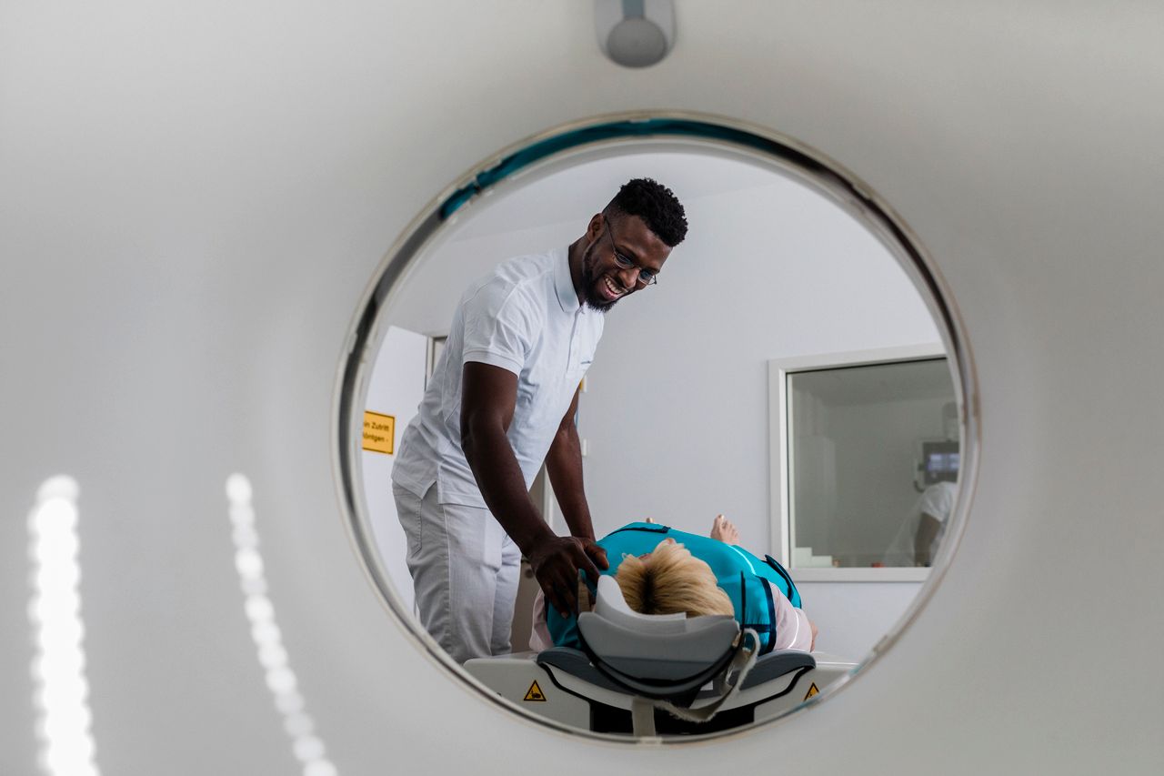
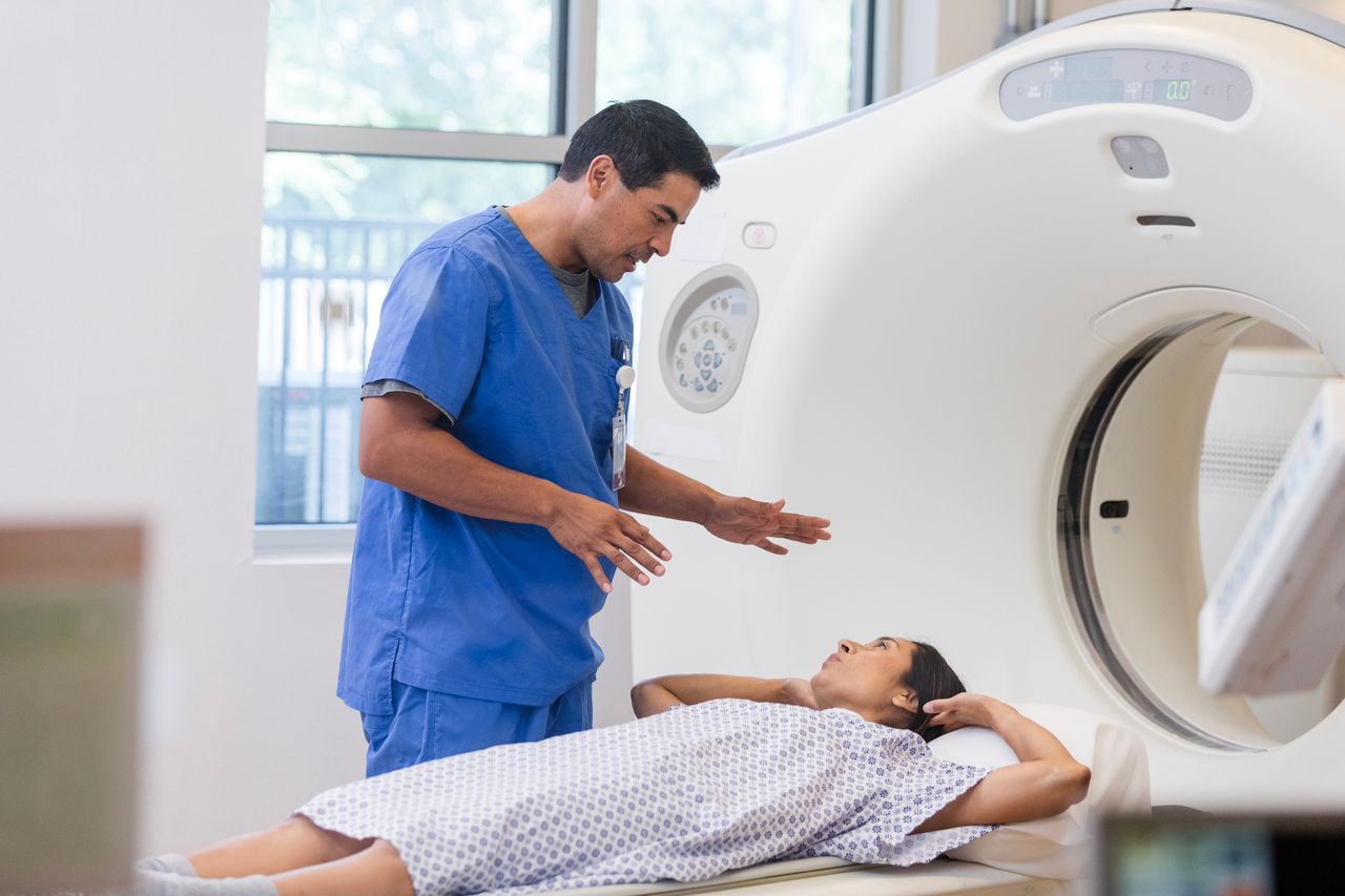
All Radiology & Imaging can be scheduled through our centralized call center and Hoag’s locations throughout Orange County offer added convenience. Our technologists and support staff are dedicated to patient-centered care. Reports are typically available within an hour of scanning, and referring physicians can choose to receive reports by fax the moment they are finalized. Critical results are always called to the referring physician immediately.
A to Z
Hoag is both a community hospital and a full service, tertiary care facility. We offer everything from basic screening tests to cutting edge procedures. Many patients are referred to Hoag from other parts of the state and country to consult with our experts. Having all of your Radiology & Imaging performed at Hoag is step toward ensuring coordinated, high-level care.
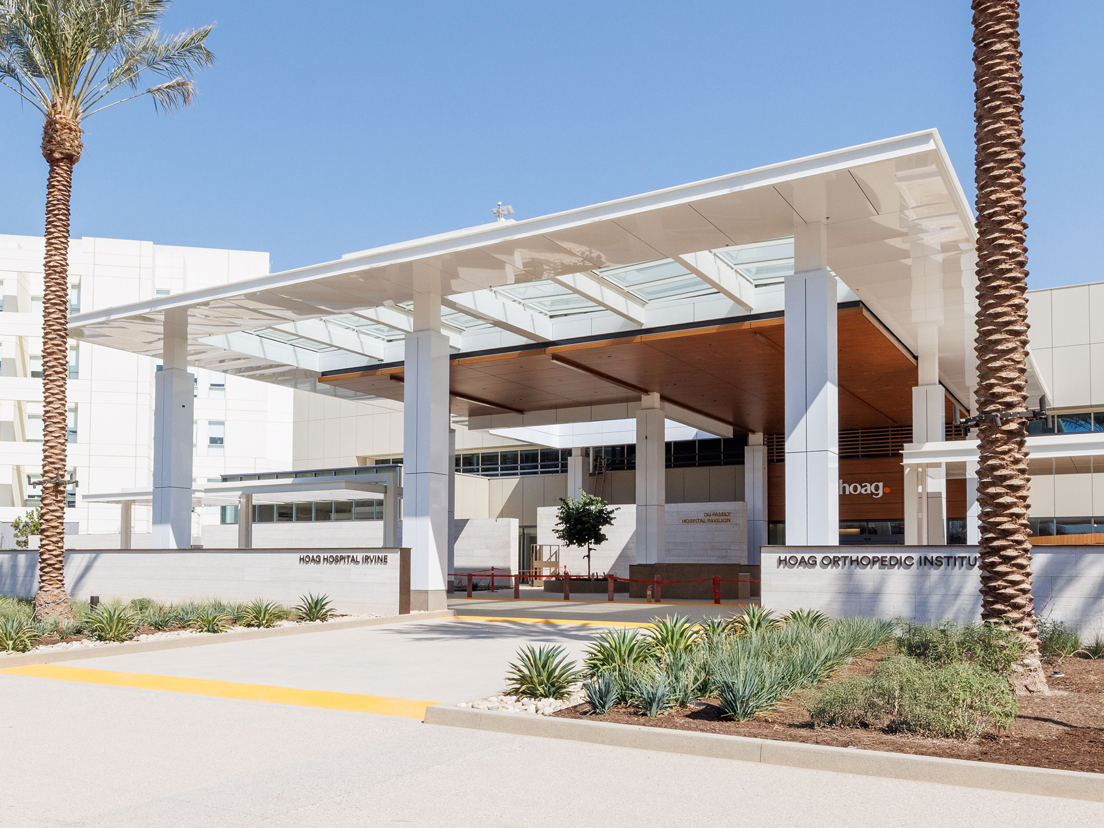
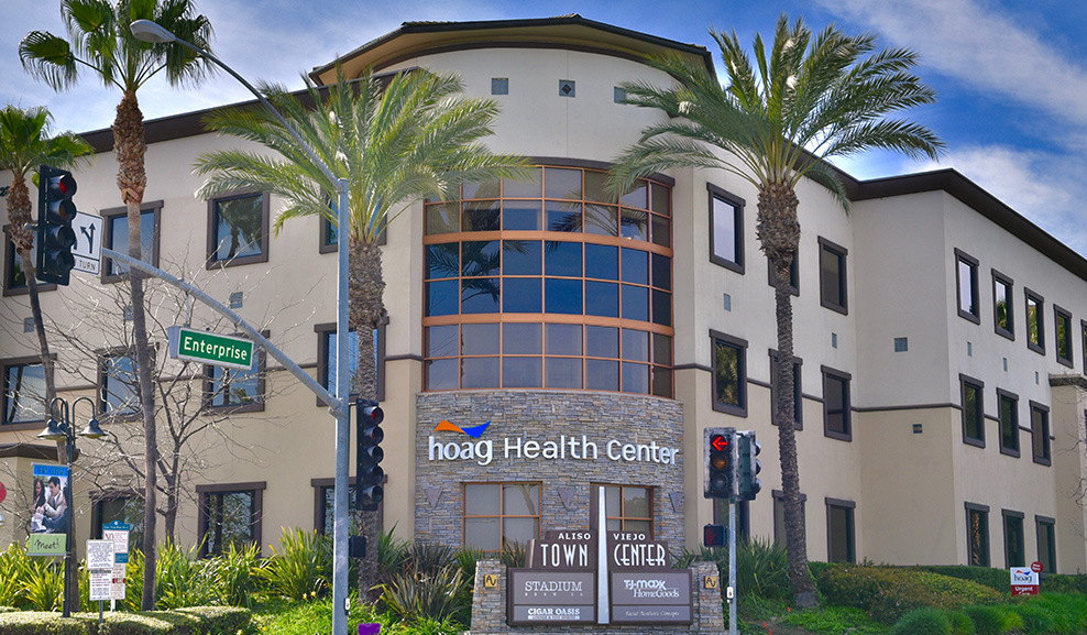
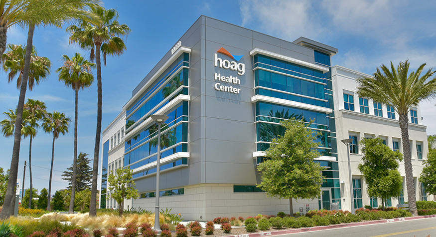
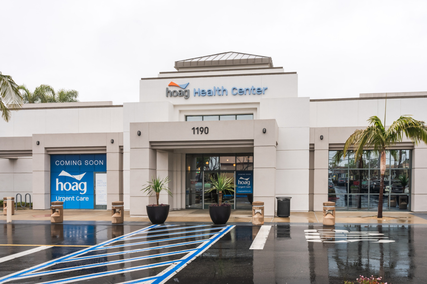
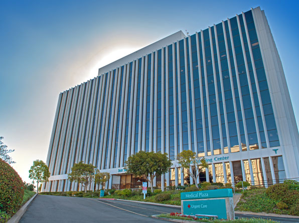
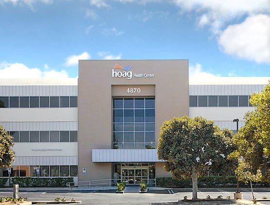
Radiology & Imaging Locations
Hoag offers radiology & imaging services at seven convenient locations throughout Orange County. Imaging services include: Advanced Musculoskeletal Imaging, Advanced Neuroimaging, Breast Biopsy, Breast MRI, Breast Ultrasound, Cardiac MRI, Computed Tomography (CT), Coronary/Cardiac CT Angiography, DEXA Scan, Interventional Neuroradiology, Interventional Radiology, Mammography, MR/PET, MRI, Nuclear Medicine, PET/CT, Spinal and Musculoskeletal Injections and Ultrasound. Services vary by location so please call ahead to confirm service offering at each location.
Radiology & Imaging Services