Gamma Knife
What is Gamma Knife?
Imagine undergoing brain surgery as an outpatient with reduced pain, a brief recovery period and no incision. Hoag Gamma Knife Center is the only facility in Orange County providing this effective treatment option for patients with brain disease.
Pickup Family Neurosciences Institute was the first center in Southern California to employ the revolutionary Leksell Gamma Knife® Perfexion™—the newest, most efficient and precise radiosurgical device available. With an expanded treatment area and enhanced accuracy, this Gamma Knife technology benefits significantly more patients who can now be treated with Gamma Knife radiosurgery instead of a more invasive procedure.
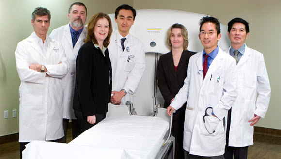
The Hoag Neurosciences Gamma Knife Team
Developed in Sweden in the 1950s, the Gamma Knife has been in use in the United States for more than two decades, providing patients with a treatment option for brain disorders that were once only treatable by open skull surgery. With unsurpassed and proven clinical outcomes, the Gamma Knife is the gold standard in cranial radiosurgery.
In Gamma Knife radiosurgery, a focused dose of radiation is used to stop and/or reduce the growth of abnormal tissue. The Gamma Knife radiation distorts the DNA mapping of the cells and renders them unable to divide. The 192 beams of radiation are focused on the abnormal tissue area with the area of intersection conforming to the size and shape of the target. Each of the individual beams provides a relatively small, harmless dose of radiation. Only at the point where the narrow beams converge is the radiation at its most powerful, therefore preventing injury to surrounding healthy tissue.
Equipped with the most advanced radiosurgical device available, Leksell Gamma Knife® Perfexion™, Hoag’s treatment system takes Gamma Knife radiosurgery one step further.
Leksell Gamma Knife Perfexion delivers the most efficient and precise radiosurgery treatments to date. With an expanded treatment area and enhanced accuracy, this sophisticated system allows Hoag clinicians to treat tumors that were unreachable using previous technology. Gamma Knife treatments take place in a single day without an incision, allowing patients to quickly resume their normal activities.
Gamma Knife Radiosurgery Benefits
Gold Standard in Cranial Radiosurgery
Gamma Knife radiosurgery is one of the most advanced means available for the treatment of brain disease. Backed by over two decades of clinical application in the U.S. and supported by thousands of peer-reviewed journal articles, no other neurosurgical tool has met with such impressive results—no other existing technology is able to deliver high levels of radiation to a tumor or other target, while sparing surrounding healthy tissue.
Minimized Health Risks
Since the Gamma Knife does not require an incision, and in most cases is used with only mild sedation and a local anesthetic, risks associated with conventional surgery are eliminated. Additionally, the design of the Gamma Knife ensures that only abnormal tissue is treated—surrounding healthy tissue being spared.
Shorter Hospitalization
Gamma Knife patients generally leave the hospital the day of treatment and are able to resume normal activities in a short period of time. Conventional neurosurgery requires lengthy hospital stays and a long recovery period.
Lowered Costs
Gamma Knife offers a cost-effective treatment option due to the minimal hospital stay and brief recovery period. Most major insurance carriers cover Gamma Knife treatment, including Medicare, Medi-Cal, most HMOs and PPOs.
Gamma Knife Radiation and Imaging Techniques at Hoag
Advanced Imaging Technique
The evolution and success of Gamma Knife Radiosurgery is directly dependent on the imaging modalities available to determine the location of the tumor or AVM. A Gamma Knife (GK) treatment center must have the accompanying state-of-the-art imaging modalities and neuro-oncologic radiology specialists in order to maximize the efficacy of the GK treatment unit and team. Hoag is fortunate to have both the technology and the staffing to collaborate on neurological and Gamma Knife patients.Learn more about the advanced Neuro Imaging available at Hoag.
Gamma Knife Radiation
The leading edge technique is an innovative process developed to treat one of the most common types of glioma, glioblastoma multiforme. This treatment combines the functionality of the Gamma Knife with the advances in diagnostic radiology technology, which include Multivoxel MR-Spectroscopy scans. This unique combination targets the radiation beyond the local tumor volume to include the potential malignant tumor path. Prior to this technological spectroscopy breakthrough, it was impossible to detect where the tumor would or might spread.
Clinical Explanation
Glioblastoma multiforme (GBM) is among the most common and devastating brain tumors affecting adults. The most successful treatment regimens for GBMs include surgical resection, radiation therapy and chemotherapy, followed by stereotactic radiosurgical boost (Gamma Knife radiosurgery) and immunotherapy. Despite aggressive therapies, the malignant nature of GBMs often results in tumor recurrence. Ninety percent of GBMs recur at the site of the original tumor. In addition to local recurrence, malignant gliomas frequently spread in predictable patterns along the white matter pathways in the brain. It is via this mechanism that long-fought battles against GBMs are often lost.
Predictable Patterns of Spread in Malignant Gliomas
Traditional radiosurgical techniques have focused solely on local tumor margins, as determined by gadolinium enhancement on MRI. However, recent data suggests that by targeting the “leading edge” of these tumors, their spread along white matter pathways can be more effectively halted. FLAIR sequences and Multivoxel MR-Spect scans can be utilized to define positive areas outside of the gadolinium T1-weighted enhancing zones. Targeting these zones with gamma knife is proving to be a successful method of blocking the path of malignant gliomas.
Gamma Knife Treatment Process
The patient is admitted to the hospital on the morning of the procedure and started on intravenous fluids. A stereotactic frame is painlessly secured to the patient’s head. This head frame is a guiding device, which ensures the Gamma Knife beams are focused exactly where the treatment is needed. Mild sedation and a local anesthetic are administered prior to head frame placement. Head frame placement usually takes fewer than 10 minutes.
Imaging studies, such as MRI, CT scan and/or angiography are performed. The stereotactic frame is displayed on the imaging studies allowing the physicians to precisely localize the target.
The team of Gamma Knife physicians utilizes three-dimensional computer imaging to develop the patient’s radiosurgical treatment plan.
When the treatment plan is complete, the patient is placed on the Gamma Knife bed. The stereotactic head frame is attached to the helmet containing 192 portals through which radiation beams are focused. The frame and the helmet prevent movement, ensuring accurate alignment during treatment.
The Gamma Knife team monitors the patient throughout the procedure. Actual treatment may last as little as 10 minutes or as long as two hours. During the procedure, the patient does not hear or feel the Gamma Knife delivering the radiation treatment.
The head frame is removed upon completion of the treatment and bandages are applied to the pin site areas. The patient is observed for approximately one hour, then discharged from the hospital.
The patient is contacted by the Gamma Knife nurse the next day for follow-up and then periodically to monitor the patient’s status.
For more information, please contact Laura Heim, MSN RN at 949-764-6077
Types of Brain Tumors Treatable by Gamma Knife Neurosurgery
Acoustic Neuroma
An acoustic neuroma is a skull-based tumor that affects the sheath of the VIII cranial nerve. In general, this is a slow growing, benign tumor. Although it is a benign tumor, this tumor presses on the nerves associated with hearing and balance. The preservation of hearing is critical in the treatment of this disease.
Brain Metastases
Metastasis to the brain may originate from various different primary tumor locations including lung, breast, skin, kidney and colon. The most common source of brain metastases in males is lung cancer and in females is breast cancer. Metastatic tumors of the brain (otherwise referred to as “Mets”) can arise anywhere in the brain. Mets can present as a single lesion (solitary metastasis) or as more than one lesion (multiple metastasis). These presentations warrant different treatment regimens—gamma knife alone or in combination with surgery, radiation therapy or chemotherapy.
Malignant Glioma
Gliomas are tumors that arise in the brain tissue itself, glial tissue (as opposed to metastatic brain tumors which arise from other body organs and travel to the brain). They are the most common type of brain tumor. There are several different types of gliomas: astrocytomas, oligodendrogliomas, ependymomas and glioblastoma multiforme.Gliomas are identified by their microscopic appearance and are graded based on their level of malignancy. As the grade of the tumor increases, the tumor becomes more aggressive.
Grade I: Pilocytic Astrocytoma, which is considered benign
Grade II: Low-Grade Astrocytomas, which includes most oligodendrogliomas and ependymomas that are often mixed, hence mixed glioma. These tumors may be malignant or benign and may also be considered grade III.
Grade III: Anaplastic Astrocytoma may be a reoccurrence of a lower grade, previously treated tumor.
Grade IV: Glioblastoma Multiforme, which is the most malignant form of astrocytomas. Each grade of tumor can be treated with the Gamma Knife.
Meningioma
Meningiomas are tumors that arise from the leptomeninges or the brain lining tissue. Meningiomas are slow growing and, in general, are benign tumors. Treatment for meningiomas may include surgery, gamma knife or both.
Pituitary Tumors
The pituitary gland is also referred to as the “master” gland. This reference is made because the pituitary gland is responsible for regulating many functions performed by the other organs in the body. The pituitary gland will secrete hormones to these organs, which communicate to the organs. The gland is divided into two parts: the anterior pituitary and the posterior pituitary. Therefore, tumors of the pituitary gland can affect the entire body.